Endosonographic Characteristics of Subepithelial Lesions of the Upper Digestive Tract: Experience of a Referral Center in Colombia
DOI:
https://doi.org/10.22516/25007440.1014Keywords:
Endoscopic ultrasound, Subepithelial lesion, GIST, Follow-upAbstract
Introduction: Subepithelial lesions (SELs), described as bulges or masses covered by healthy-looking mucosa, are usually found incidentally during endoscopic studies. They are typically asymptomatic and are estimated to be identified in 1% of esophagogastroduodenoscopies performed.
Materials and methods: A descriptive study was conducted with retrospective data collection. We included all patients treated at the Unión de Cirujanos, a referral gastroenterology unit of the Coffee Region in Manizales, between January 2020 and January 2022, who underwent endoscopic ultrasonography to study subepithelial-looking lesions located in the esophagus, stomach, and duodenum.
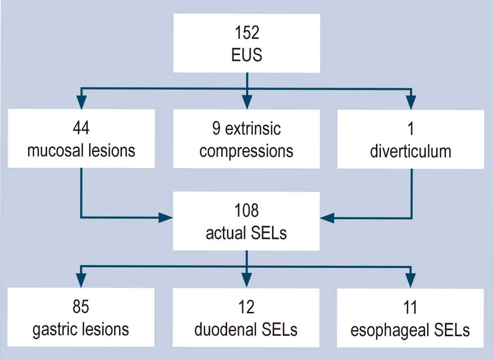
Results: 152 endoscopic ultrasounds were performed, finding 108 SELs; 66.6% of the patients were women, and the average age was 58. Most SELs were located in the stomach (78.7%), the antrum being the most frequent location. The average diameter of the gastric SELs was 14.6 mm, and 47% of the lesions depended on the fourth echolayer; the most frequent presumptive diagnoses were gastrointestinal stromal tumor (GIST; 65.8%) and lipoma (11.7%).
Conclusions: SELs of the GI tract originate in the muscularis mucosae, submucosa, or muscularis propria. They are most frequently located in the stomach, and their characterization usually requires endoscopic ultrasonography and histopathology. Treatment of these lesions remains controversial due to their low frequency, histological variety, and low malignant potential.
Downloads
References
Attila T, Aydin Ö. Lesion size determines diagnostic yield of EUS-FNA with onsite cytopathologic evaluation for upper gastrointestinal subepithelial lesions. Turkish J Gastroenterol. 2018;29(4):436-41. https://doi.org/10.5152/tjg.2018.17876
Menon L, Buscaglia JM. Endoscopic approach to subepithelial lesions. Therap Adv Gastroenterol. 2014;7(3):123-30. https://doi.org/10.1177/1756283X13513538
Nishida T, Kawai N, Yamaguchi S, Nishida Y. Submucosal tumors: Comprehensive guide for the diagnosis and therapy of gastrointestinal submucosal tumors. Dig Endosc. 2013;25(5):479-89. https://doi.org/10.1111/den.12149
Cho JW. Current guidelines in the management of upper gastrointestinal subepithelial tumors. Clin Endosc. 2016;49(3):235-40. https://doi.org/10.5946/ce.2015.096
Deprez PH, Moons L, O’toole D, Gincul R, Seisean A, Pimentel-Nunes P, et al. Endoscopic management of subepithelial lesions including neuroendocrine neoplasms: European Society of Gastro- intestinal Endoscopy (ESGE) Guideline. Endoscopy. 2022;54(4):412-429. https://doi.org/10.1055/a-1751-5742
Franco MC. Opinion: How to manage subepithelial lesions of the upper gastrointestinal tract? World J Gastrointest Endosc. 2015;7(18):1262. https://doi.org/10.4253/wjge.v7.i18.1262
Eckardt AJ, Jenssen C. Current endoscopic ultrasound-guided approach to incidental subepithelial lesions: Optimal or optional? Ann Gastroenterol. 2015;28(2):160-72.
Arango L, Sánchez A, Bautista I, Díaz C. Endosonographic Findings of Gastroduodenal Ectopic Pancreas: Experience of A Reference Center in Colombia and Literature Review. J Gastroenterol Hepatol. 2021;21(5):1-7. https://doi.org/04.2021/1.1010
Landi B, Palazzo L. The role of endosonography in submucosal tumours. Best Pract Res Clin Gastroenterol. 2009;23(5):679-701. https://doi.org/10.1016/j.bpg.2009.05.009
Hu ML, Wu KL, Changchien CS, Chuah SK, Chiu YC. Endosonographic surveillance of 1-3 cm gastric submucosal tumors originating from muscularis propria. World J Gastroenterol. 2017;23(12):2194-200. https://doi.org/10.3748/wjg.v23.i12.2194
Park EY, Kim GH. Diagnosis of gastric subepithelial tumors using endoscopic ultrasonography or abdominopelvic computed tomography: Which is better? Clin Endosc. 2019;52(6):519-20. https://doi.org/10.5946/ce.2019.188
Hohenberger P, Raut CP, Rutkowski P. Gastrointestinal stromal tumors. Visc Med. 2018;34(5):332-3. https://doi.org/10.1159/000494077
Sanaei O, Fernández-Esparrach G, De La Serna-Higuera C, Carrara S, Kumbhari V, El Zein MH, et al. EUS-guided 22-gauge fine needle biopsy versus single-incision with needle knife for the diagnosis of upper gastrointestinal subepithelial lesions: a randomized controlled trial. Endosc Int Open. 2020;08(03):E266-73. https://doi.org/10.1055/a-1075-1900
Dhaliwal A, Kolli S, Dhindsa BS, Devani K, Ramai D, Sayles H, et al. Clinical efficacy and safety of mucosal incision-assisted biopsy for the diagnosis of upper gastrointestinal subepithelial tumors: A systematic review and meta-analysis. Ann Gastroenterol. 2020;33(2):155-61. https://doi.org/10.20524/aog.2020.0460
de Moura DTH, McCarty TR, Jirapinyo P, Ribeiro IB, Flumignan VK, Najdawai F, et al. EUS-guided fine-needle biopsy sampling versus FNA in the diagnosis of subepithelial lesions: a large multicenter study. Gastrointest Endosc. 2020;92(1):108-119.e3. https://doi.org/10.1016/j.gie.2020.02.021
Trindade AJ, Benias PC, Alshelleh M, Bazarbashi AN, Tharian B, Inamdar S, et al. Fine-needle biopsy is superior to fine-needle aspiration of suspected gastrointestinal stromal tumors: a large multicenter study. Endosc Int Open. 2019;07(07):E931-6. https://doi.org/10.1055/a-0953-1640
Kim GH, Cho YK, Kim EY, Kim HK, Cho JW, Lee TH, et al. Comparison of 22-gauge aspiration needle with 22-gauge biopsy needle in endoscopic ultrasonography-guided subepithelial tumor sampling. Scand J Gastroenterol. 2014;49(3):347-54. https://doi.org/10.3109/00365521.2013.867361
Oberg K, Couvelard A, Delle Fave G, Gross D, Grossman A, Jensen RT, et al. ENETS Consensus Guidelines for the Standards of Care in Neuroendocrine Tumors: Biochemical Markers. Neuroendocrinology. 2017;105(3):201-11. https://doi.org/10.1159/000472254
Esmo T, Sarcoma E, Working N. Gastrointestinal stromal tumours: ESMO Clinical Practice Guidelines for diagnosis, treatment and follow-up. Ann Oncol. 2014;25(Suppl 3):iii21-6. https://doi.org/10.1093/annonc/mdu255
Landi B, Blay JY, Bonvalot S, Brasseur M, Coindre JM, Emile JF, et al. Gastrointestinal stromal tumours (GISTs): French Intergroup Clinical Practice Guidelines for diagnosis, treatments and follow-up (SNFGE, FFCD, GERCOR, UNICANCER, SFCD, SFED, SFRO). Dig Liver Dis. 2019;51(9):1223-31. https://doi.org/10.1016/j.dld.2019.07.006
Dumonceau JM, Deprez PH, Jenssen C, Iglesias-Garcia J, Larghi A, Vanbiervliet G, et al. Indications, results, and clinical impact of endoscopic ultrasound (EUS)-guided sampling in gastroenterology: European Society of Gastrointestinal Endoscopy (ESGE) Clinical Guideline - Updated January 2017. Endoscopy. 2017;49(7):695-714. https://doi.org/10.1055/s-0043-109021
Kumar S, Chandrasekhara V, Kochman ML, Ahmad N, Attalla S, Ho IK, et al. Ligation-assisted endoscopic mucosal resection for esophageal granular cell tumors is safe and effective. Dis Esophagus. 2020;33(8):1-5. https://doi.org/10.1093/dote/doaa027
Peng W, Tan S, Huang S, Ren Y, Li H, Peng Y, et al. Efficacy and safety of submucosal tunneling endoscopic resection for upper gastrointestinal submucosal tumors with more than 1-year’ follow-up: a systematic review and meta-analysis. Scand J Gastroenterol. 2019;54(4):397-406. https://doi.org/10.1080/00365521.2019.1591500
Downloads
Published
How to Cite
Issue
Section
License
Copyright (c) 2023 Revista colombiana de Gastroenterología

This work is licensed under a Creative Commons Attribution-NonCommercial-NoDerivatives 4.0 International License.
Aquellos autores/as que tengan publicaciones con esta revista, aceptan los términos siguientes:
Los autores/as ceden sus derechos de autor y garantizarán a la revista el derecho de primera publicación de su obra, el cuál estará simultáneamente sujeto a la Licencia de reconocimiento de Creative Commons que permite a terceros compartir la obra siempre que se indique su autor y su primera publicación en esta revista.
Los contenidos están protegidos bajo una licencia de Creative Commons Reconocimiento-NoComercial-SinObraDerivada 4.0 Internacional.

| Article metrics | |
|---|---|
| Abstract views | |
| Galley vies | |
| PDF Views | |
| HTML views | |
| Other views | |