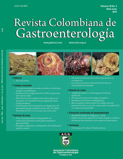
Ecoendoscopia en la evaluación de las lesiones subepiteliales duodenales
DOI:
https://doi.org/10.22516/25007440.41Palavras-chave:
Subepitelial, duodeno, ecoendoscopia, GIST, neuroendocrinoResumo
Las lesiones o tumores subepiteliales (TSE) son raros. Se considera que en 1 de cada 300 endoscopias puede encontrarse un TSE, lo que corresponde al 0,36% de las endoscopias. De estos, solo el 10% se ubican en el duodeno, lo que proporciona una idea de lo infrecuente que es este hallazgo. Al realizar la endoscopia y detectar un TSE en el duodeno, inicialmente se describirá su tamaño, forma, color, movilidad, pulsación y finalmente la consistencia, la cual puede evaluarse con la pinza de biopsia cerrada. Esto permitirá detectar si es quístico, firme o si tiene el “signo de la almohada”, que es altamente sugestivo de lipoma. En caso contrario, está indicada la realización de una ecoendoscopia, especialmente si el tumor es mayor de 1 cm.
Downloads
Referências
Sonnenberg A, Amorosi SL, Lacey MJ, et al. Patterns of endoscopy in the United States: analysis of data from the Centers for Medicare and Medicaid Services and the National Endoscopic Database. Gastrointest Endosc. 2008;67(3):489-96.
Park CH, Kim B, Chung H, Lee H, Park JC, Shin SK, et al. Endoscopic quality indicators for esophagogastro- duodenoscopy in gastric cancer screening. Dig Dis Sci. 2015;60(1):38-46.
Buscaglia J, Nagula S, Jayaraman V, Robbins D, et al. Diagnostic yield and safety of jumbo biopsy forceps in patients with subepithelial lesions of the upper and lower GI tract. Gastrointest Endosc. 2012;75:1147-52.
Papanikolaou IS, Triantafyllou K, Kourikou A, et al. Endoscopic ultrasonography for gastric submucosal lesions. World J Gastrointest Endosc. 2011;3(5):86-94.
Muenst S, Thies S, Went P, et al. Frequency, phenotype, and genotype of minute gastrointestinal stromal tumors in the sto- mach: an autopsy study. Hum Pathol. 2011;42(12):1849-54.
Jenssen C, Dietrich CF. Endoscopic ultrasound in sube- pithelial tumors of the gastrointestinal tract. En: Dietrich CF, editor. Endoscopic ultrasound: an introductory manual and atlas. New York: Thieme; 2006. p. 121-54.
Souquet JC, Bobichon R. Role of endoscopic ultrasound in the management of submucosal tumours in the esophagus and stomach. Acta Endoscop. 1996;26:307-12.
Hwang JH, Saunders MD, Rulyak SJ, et al. A prospective study comparing endoscopy and EUS in the evaluation of GI sube- pithelial masses. Gastrointest Endosc. 2005;62(2):202-8.
Boyce GA, Sivak Jr. MV, Rosch T, et al. Evaluation of sub- mucosal upper gastrointestinal tract lesions by endoscopic ultrasound. Gastrointest Endosc. 1991;37:449-54.
Caletti G, Zani L, Bolondi L, et al. Endoscopic ultraso- nography in the diagnosis of gastric submucosal tumor. Gastrointest Endosc. 1989;35:413-8.
Bruno M, Carucci P, Repici A, Pellicano R, Mezzabotta L, Goss M, et al. The natural history of gastrointestinal subepithelial tumors arising from muscularis propria: an endoscopic ultrasound survey. J Clin Gastroenterol.
;43(9):821-5.
Soga J. Early-stage carcinoids of the gastrointestinal tract: an analysis of 1914 reported cases. Cancer. 2005;103:1587-95. 13. Karaca C, Turner BG, Cizginer S, Forcione D, Brugge W. Accuracy of EUS in the evaluation of small gastric subepithelial lesions. Gastrointest Endosc. 2010;71(4):722-7. 14. Sepe PS, Moparty B, Pitman MB, Saltzman JR, Brugge WR. EUS-guided FNA for the diagnosis of GI stromal cell tumors: sensitivity and cytologic yield. Gastrointest Endosc. 2009;70(2):254-61.
Mekky MA, Yamao K, Sawaki A, et al. Diagnostic utility of EUS-guided FNA in patients with gastric submucosal tumors. Gastrointest Endosc. 2010;71:913-9.
Hoda KM, Rodriquez SA, Faigel DO. EUS-guided sam- pling of suspected GI stromal tumors. Gastrointest Endosc. 2009;69:1218-23.
Sepe PS, Moparty B, Pitman MB, et al. EUS-guided FNA for the diagnosis of GI stromal cell tumors: sensitivity and cytologic yield. Gastrointest Endosc. 2009;70:254-61.
Larghi A, Fuccio L, Chiarello G, Attili F, Vanella G, Paliani GB, et al. Fine-needle tissue acquisition from subepithelial lesions using a forward-viewing linear echoendoscope: Endoscopy. 2014;46(1):39-45.
Hamada T, Yasunaga H, Nakai Y, Isayama H, Horiguchi H,
Matsuda S, et al. Rarity of severe bleeding and perforation in endoscopic ultrasound-guided fine needle aspiration for submucosal tumors. Dig Dis Sci. 2013;58(9):2634-8.
Waisberg J, Joppert-Netto G, Vasconcellos C, Sartini GH, Miranda LS, Franco MI. Carcinoid tumor of the duodenum: a rare tumor at an unusual site. Case series from a single ins- titution. Arq Gastroenterol. 2013;50(1):3-9.
Tai WP, Yue H. Endoscopic mucosa resection of a duode- num carcinoid tumor of 1.2 cm diameter: a case report. Med Oncol. 2009;26:319-21.
Tsujimoto H, Ichikura T, Nagao S, Sato T, Ono S, Aiko S, et al. Minimally invasive surgery for resection of duodenal carcinoid tumors: endoscopic full-thickness resection under laparoscopic observation. Surg Endosc. 2010;24:471-5.
Zyromski NJ, Kendrick ML, Nagorney DM, Grant CS, Donohue JH, Farnell MB, et al. Duodenal carcinoid tumors: how aggressive should we be? J Gastrointest Surg. 2001;5:588-93.
IorioN,SawayaRA,FriedenbergFK.Reviewarticle:thebio- logy, diagnosis and management of gastrointestinal stromal tumours. Aliment Pharmacol Ther. 2014;39(12):1376-86.
Chok AY, Koh YX, Ow MY, Allen JC Jr, Goh BK. A systema- tic review and meta-analysis comparing pancreaticoduode- nectomy versus limited resection for duodenal gastrointesti- nal stromal tumors. Ann Surg Oncol. 2014;21(11)3429-38.
Matsumoto S, Miyatani H, Yoshida Y, Nokubi M. Duodenal carcinoid tumors: 5 cases treated by endoscopic submucosal dissection. Gastrointest Endosc. 2011;74:1152-6.
Huang ZG, Zhang XS, Huang SL, Yuan XG. Endoscopy dis- section of small stromal tumors emerged from the muscu- laris propria in the upper gastrointestinal tract: Preliminary study. World J Gastrointest Endosc. 2012;4(12):565-70.
Downloads
Publicado
Como Citar
Edição
Seção
Licença
Aquellos autores/as que tengan publicaciones con esta revista, aceptan los términos siguientes:
Los autores/as ceden sus derechos de autor y garantizarán a la revista el derecho de primera publicación de su obra, el cuál estará simultáneamente sujeto a la Licencia de reconocimiento de Creative Commons que permite a terceros compartir la obra siempre que se indique su autor y su primera publicación en esta revista.
Los contenidos están protegidos bajo una licencia de Creative Commons Reconocimiento-NoComercial-SinObraDerivada 4.0 Internacional.


| Métricas do artigo | |
|---|---|
| Vistas abstratas | |
| Visualizações da cozinha | |
| Visualizações de PDF | |
| Visualizações em HTML | |
| Outras visualizações | |


