Utilidad de la elastografía cuantitativa por ultrasonografía endoscópica (USE), para el diagnóstico de las lesiones sólidas del páncreas (LSP).
DOI:
https://doi.org/10.22516/25007440.643Palavras-chave:
Elastografía, ultrasonido endoscópico, lesión pancreática s´lida, elasticidad, páncreasResumo
Introducción: la punción con aguja fina guiada por ultrasonografía endoscópica (PAF-USE) permite un diagnóstico de las lesiones sólidas del páncreas (LSP) con una sensibilidad de alrededor del 85 % en la literatura mundial y aún más baja en nuestro medio, por lo cual se requiere explorar nuevos accesorios (agujas) o técnicas tales como la elastografía, que mejoren esta sensibilidad. Esta última permite la cuantificación de la rigidez del tejido con altos grados de precisión y desde 2001 se ha aplicado al diagnóstico de tumores sólidos de diversos órganos como mama y tiroides, músculo, entre otros; y desde 2006 se ha empleado para las LSP y ha demostrado su utilidad como complemento a las herramientas diagnósticas disponibles, ya que mejora la precisión de la biopsia por PAF-USE al seleccionar el área más sospechosa para ser puncionada y también guía el manejo clínico cuando la PAF-USE es negativa o no concluyente.
Objetivo: evaluar el rendimiento diagnóstico de la elastografía cuantitativa de strain ratio (SR) obtenida por ecoendoscopia en las lesiones sólidas pancreáticas teniendo como patrón de oro el diagnóstico citopatológico.
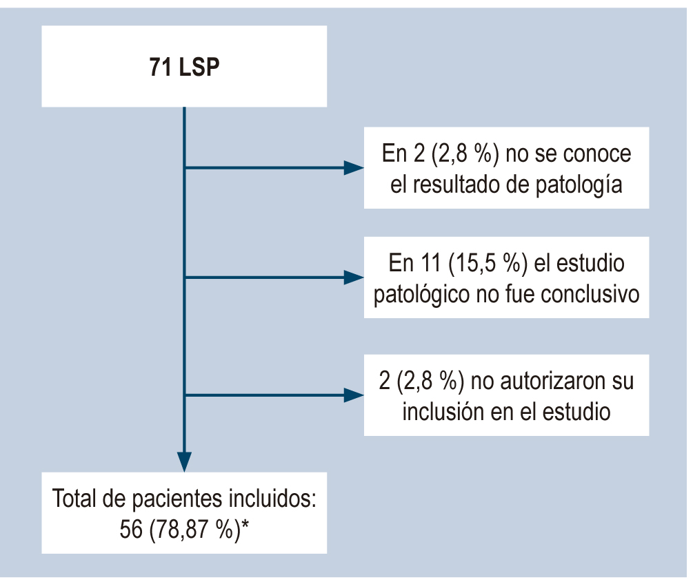
Métodos: 71 pacientes (rango de edad: 35-89, media: 62,2 años); de estos, 35 mujeres fueron sometidas a USE para la evaluación de LSP. El diseño del estudio fue de corte transversal, prospectivo y de un solo centro. La USE se realizó con un ecoendoscopio Pentax lineal y un procesador Hitachi-Noblus. La lesión (área A) y un área de referencia B se seleccionaron para calcular la relación de deformación (B/A, SR expresada en %). Se tomó como punto de corte SR para definir las lesiones malignas (duras) SR > 22 teniendo en cuenta la evidencia actualmente disponible; estos resultados se compararon con la citopatología de las muestras obtenidas por punción guiada por USE. Después de la aplicación de criterios de exclusión, se realiza el análisis estadístico de 56 pacientes y se considera el valor p < 0,05. Se calculó la sensibilidad, especificidad, valor predictivo positivo (VPP), valor predictivo negativo (VPN) y precisión diagnóstica comparando la elastografía SR con los diagnósticos finales por citopatología.
Resultados: la elastografía cuantitativa SR (%) permite detectar las LSP malignas con sensibilidad del 94,6 % (intervalo de confianza [IC] del 95 %: 85,4 %-98,2 %), especificidad del 89,3 % (IC 95 %: 78,5 %-95,0 %), VPP del 89,8 % (IC 95 %: 79,5 %-95,3 %); VPN del 94,3 % (IC 95 %: 84,6 %-98,1 %) y exactitud del 92,0 % (IC 95 %: 85,4 %-95,7 %).
Conclusión: la elastografía cuantitativa SR por USE en LSP es un complemento útil que mejora la precisión de la PAF-USE al seleccionar el área más sospechosa para ser puncionada y guiar el manejo clínico cuando la PAF-USE es negativa o no concluyente, ya que tiene una alta sensibilidad y especificad en el diagnóstico de las LSP malignas.
Downloads
Referências
Iglesias-García J, Lariño-Noia J, Domínguez-Muñoz JE. New Imaging Techniques: Endoscopic Ultrasound-Guided Elastography. Gastrointest Endosc Clin N Am. 2017;27(4):551-567. https://doi.org/10.1016/j.giec.2017.06.001
Luthra AK, Evans JA. Review of current and evolving clinical indications for endoscopic ultrasound. World J Gastrointest Endosc. 2016;8(3):157-64. https://doi.org/10.4253/wjge.v8.i3.157
Kleeff J, Korc M, Apte M, La Vecchia C, Johnson CD, Biankin AV, Neale RE, Tempero M, Tuveson DA, Hruban RH, Neoptolemos JP. Pancreatic cancer. Nat Rev Dis Primers. 2016;2:16022. https://doi.org/10.1038/nrdp.2016.22
Bray F, Ferlay J, Soerjomataram I, Siegel RL, Torre LA, Jemal A. Global cancer statistics 2018: GLOBOCAN estimates of incidence and mortality worldwide for 36 cancers in 185 countries. CA Cancer J Clin. 2018;68(6):394-424. https://doi.org/10.3322/caac.21492
Xiao AY, Tan ML, Wu LM, Asrani VM, Windsor JA, Yadav D, Petrov MS. Global incidence and mortality of pancreatic diseases: a systematic review, meta-analysis, and meta-regression of population-based cohort studies. Lancet Gastroenterol Hepatol. 2016;1(1):45-55. https://doi.org/10.1016/S2468-1253(16)30004-8
Wang W, Shpaner A, Krishna SG, Ross WA, Bhutani MS, Tamm EP, Raju GS, Xiao L, Wolff RA, Fleming JB, Lee JH. Use of EUS-FNA in diagnosing pancreatic neoplasm without a definitive mass on CT. Gastrointest Endosc. 2013;78(1):73-80. https://doi.org/10.1016/j.gie.2013.01.040
Hewitt MJ, McPhail MJ, Possamai L, Dhar A, Vlavianos P, Monahan KJ. EUS-guided FNA for diagnosis of solid pancreatic neoplasms: a meta-analysis. Gastrointest Endosc. 2012;75(2):319-31. https://doi.org/10.1016/j.gie.2011.08.049
Bhatia V, Varadarajulu S. Endoscopic ultrasonography-guided tissue acquisition: How to achieve excellence. Dig Endosc. 2017;29(4):417-430. https://doi.org/10.1111/den.12823
Polkowski M, Larghi A, Weynand B, Boustière C, Giovannini M, Pujol B, Dumonceau JM; European Society of Gastrointestinal Endoscopy (ESGE). Learning, techniques, and complications of endoscopic ultrasound (EUS)-guided sampling in gastroenterology: European Society of Gastrointestinal Endoscopy (ESGE) Technical Guideline. Endoscopy. 2012;44(2):190-206. https://doi.org/10.1055/s-0031-1291543
Korenblit J, Tholey DM, Tolin J, Loren D, Kowalski T, Adler DG, Davolos J, Siddiqui AA. Effect of the time of day and queue position in the endoscopic schedule on the performance characteristics of endoscopic ultrasound-guided fine-needle aspiration for diagnosing pancreatic malignancies. Endosc Ultrasound. 2016;5(2):78-84. https://doi.org/10.4103/2303-9027.180470
Ramesh J, Bang JY, Hebert-Magee S, Trevino J, Eltoum I, Frost A, Hasan MK, Logue A, Hawes R, Varadarajulu S. Randomized Trial Comparing the Flexible 19G and 25G Needles for Endoscopic Ultrasound-Guided Fine Needle Aspiration of Solid Pancreatic Mass Lesions. Pancreas. 2015;44(1):128-33. https://doi.org/10.1097/MPA.0000000000000217
Madhoun MF, Wani SB, Rastogi A, Early D, Gaddam S, Tierney WM, Maple JT. The diagnostic accuracy of 22-gauge and 25-gauge needles in endoscopic ultrasound-guided fine needle aspiration of solid pancreatic lesions: a meta-analysis. Endoscopy. 2013;45(2):86-92. https://doi.org/10.1055/s-0032-1325992
Kamata K, Kitano M, Yasukawa S, Kudo M, Chiba Y, Ogura T, Higuchi K, Fukutake N, Ashida R, Yamasaki T, Nebiki H, Hirose S, Hoki N, Asada M, Yazumi S, Takaoka M, Okazaki K, Matsuda F, Okabe Y, Yanagisawa A. Histologic diagnosis of pancreatic masses using 25-gauge endoscopic ultrasound needles with and without a core trap: a multicenter randomized trial. Endoscopy. 2016;48(7):632-8. https://doi.org/10.1055/s-0042-106294
Nakai Y, Isayama H, Chang KJ, Yamamoto N, Hamada T, Uchino R, Mizuno S, Miyabayashi K, Yamamoto K, Kawakubo K, Kogure H, Sasaki T, Hirano K, Tanaka M, Tada M, Fukayama M, Koike K. Slow pull versus suction in endoscopic ultrasound-guided fine-needle aspiration of pancreatic solid masses. Dig Dis Sci. 2014;59(7):1578-85. https://doi.org/10.1007/s10620-013-3019-9
Bang JY, Magee SH, Ramesh J, Trevino JM, Varadarajulu S. Randomized trial comparing fanning with standard technique for endoscopic ultrasound-guided fine-needle aspiration of solid pancreatic mass lesions. Endoscopy. 2013;45(6):445-50. https://doi.org/10.1055/s-0032-1326268
Suzuki R, Irisawa A, Bhutani MS, Hikichi T, Takagi T, Sato A, Sato M, Ikeda T, Watanabe K, Nakamura J, Tasaki K, Obara K, Ohira H. Prospective evaluation of the optimal number of 25-gauge needle passes for endoscopic ultrasound-guided fine-needle aspiration biopsy of solid pancreatic lesions in the absence of an onsite cytopathologist. Dig Endosc. 2012;24(6):452-6. https://doi.org/10.1111/j.1443-1661.2012.01311.x
Hébert-Magee S, Bae S, Varadarajulu S, Ramesh J, Frost AR, Eloubeidi MA, Eltoum IA. The presence of a cytopathologist increases the diagnostic accuracy of endoscopic ultrasound-guided fine needle aspiration cytology for pancreatic adenocarcinoma: a meta-analysis. Cytopathology. 2013;24(3):159-71. https://doi.org/10.1111/cyt.12071
Cui XW, Chang JM, Kan QC, Chiorean L, Ignee A, Dietrich CF. Endoscopic ultrasound elastography: Current status and future perspectives. World J Gastroenterol. 2015;21(47):13212-24. https://doi.org/10.3748/wjg.v21.i47.13212
Ueno E, Tohno E, Soeda S, Asaoka Y, Itoh K, Bamber JC, Blaszçzyk M, Davey J, Mckinna JA. Dynamic tests in real-time breast echography. Ultrasound Med Biol. 1988;14 Suppl 1:53-7. https://doi.org/10.1016/0301-5629(88)90047-6
Ophir J, Céspedes I, Ponnekanti H, Yazdi Y, Li X. Elastography: a quantitative method for imaging the elasticity of biological tissues. Ultrason Imaging. 1991;13(2):111-34. https://doi.org/10.1177/016173469101300201
Chantarojanasiri T, Kongkam P. Endoscopic ultrasound elastography for solid pancreatic lesions. World J Gastrointest Endosc. 2017;9(10):506-513. https://doi.org/10.4253/wjge.v9.i10.506
Shiina T, Nitta N, Ueno E, Bamber JC. Real time tissue elasticity imaging using the combined autocorrelation method. J Med Ultrason (2001). 2002;29(3):119-28. https://doi.org/10.1007/BF02481234
Giovannini M, Hookey LC, Bories E, Pesenti C, Monges G, Delpero JR. Endoscopic ultrasound elastography: the first step towards virtual biopsy? Preliminary results in 49 patients. Endoscopy. 2006;38(4):344-8. https://doi.org/10.1055/s-2006-925158
Gennisson JL, Deffieux T, Fink M, Tanter M. Ultrasound elastography: principles and techniques. Diagn Interv Imaging. 2013;94(5):487-95. https://doi.org/10.1016/j.diii.2013.01.022
Costache MI, Dumitrescu D, Săftoiu A. Technique of qualitative and semiquantitative EUS elastography in pancreatic examination. Endosc Ultrasound. 2017;6(Suppl 3):S111-S114. https://doi.org/10.4103/eus.eus_75_17
Declaración de Helsinki de la AMM. Principios éticos para las investigaciones médicas en seres humanos [internet]. AMM [consultado el 10 de agosto de 2019]. Disponible en: www.wma.net/es/policies-post/declaracion-de-helsinki-de-la-amm-principios-eticos-para-las-investigaciones-medicas-en-seres-humanos
Pérez Cruz PE, Acevedo F. Escalas de estado funcional (o performance status) en cáncer. Gastroenterol Latinoam. 2014;25(3):219-226.
Wani S, Wallace MB, Cohen J, Pike IM, Adler DG, Kochman ML, Lieb JG 2nd, Park WG, Rizk MK, Sawhney MS, Shaheen NJ, Tokar JL. Quality indicators for EUS. Am J Gastroenterol. 2015;110(1):102-13. https://doi.org/10.1038/ajg.2014.387
Villa NA, Berzosa M, Wallace MB, Raijman I. Endoscopic ultrasound-guided fine needle aspiration: The wet suction technique. Endosc Ultrasound. 2016;5(1):17-20. https://doi.org/10.4103/2303-9027.175877
Polkowski M, Jenssen C, Kaye P, Carrara S, Deprez P, Gines A, Fernández-Esparrach G, Eisendrath P, Aithal GP, Arcidiacono P, Barthet M, Bastos P, Fornelli A, Napoleon B, Iglesias-Garcia J, Seicean A, Larghi A, Hassan C, van Hooft JE, Dumonceau JM. Technical aspects of endoscopic ultrasound (EUS)-guided sampling in gastroenterology: European Society of Gastrointestinal Endoscopy (ESGE) Technical Guideline - March 2017. Endoscopy. 2017;49(10):989-1006. https://doi.org/10.1055/s-0043-119219
Attam R, Arain MA, Bloechl SJ, Trikudanathan G, Munigala S, Bakman Y, Singh M, Wallace T, Henderson JB, Catalano MF, Guda NM. “Wet suction technique (WEST)”: a novel way to enhance the quality of EUS-FNA aspirate. Results of a prospective, single-blind, randomized, controlled trial using a 22-gauge needle for EUS-FNA of solid lesions. Gastrointest Endosc. 2015;81(6):1401-7. https://doi.org/10.1016/j.gie.2014.11.023
Lee JK, Choi JH, Lee KH, Kim KM, Shin JU, Lee JK, Lee KT, Jang KT. A prospective, comparative trial to optimize sampling techniques in EUS-guided FNA of solid pancreatic masses. Gastrointest Endosc. 2013;77(5):745-51. https://doi.org/10.1016/j.gie.2012.12.009
Dietrich CF, Bibby E, Jenssen C, Saftoiu A, Iglesias-Garcia J, Havre RF. EUS elastography: How to do it? Endosc Ultrasound. 2018;7(1):20-28. https://doi.org/10.4103/eus.eus_49_17
Dietrich CF, Săftoiu A, Jenssen C. Real time elastography endoscopic ultrasound (RTE-EUS), a comprehensive review. Eur J Radiol. 2014;83(3):405-14. https://doi.org/10.1016/j.ejrad.2013.03.023
Kamata K, Kitano M, Omoto S, Kadosaka K, Miyata T, Minaga K, Yamao K, Imai H, Kudo M. New endoscopic ultrasonography techniques for pancreaticobiliary diseases. Ultrasonography. 2016;35(3):169-79. https://doi.org/10.14366/usg.15042
Kim SY, Cho JH, Kim YJ, Kim EJ, Park JY, Jeon TJ, Kim YS. Diagnostic efficacy of quantitative endoscopic ultrasound elastography for differentiating pancreatic disease. J Gastroenterol Hepatol. 2017;32(5):1115-1122. https://doi.org/10.1111/jgh.13649
Itokawa F, Itoi T, Sofuni A, Kurihara T, Tsuchiya T, Ishii K, Tsuji S, Ikeuchi N, Umeda J, Tanaka R, Yokoyama N, Moriyasu F, Kasuya K, Nagao T, Kamisawa T, Tsuchida A. EUS elastography combined with the strain ratio of tissue elasticity for diagnosis of solid pancreatic masses. J Gastroenterol. 2011;46(6):843-53. https://doi.org/10.1007/s00535-011-0399-5
Strang AM, Lockhart ME, Kenney PJ, Amling CL, Urban DA, El-Galley R, Burns JR, Colli JL, Hammontree LN, Kolettis PN. Computerized tomographic angiography for renal donor evaluation leads to a higher exclusion rate. J Urol. 2007;177(5):1826-9. https://doi.org/10.1016/j.juro.2007.01.007
Pitts A, Nissen NN, Waxman A, Yu R. Unsuspected fluorodeoxyglucose positron emission tomography (FDG-PET)-positive pancreatic lesions: prevalence and significance. Pancreas. 2013;42(7):1191-3. https://doi.org/10.1097/MPA.0b013e318287d06e
Weckesser M, Schober O. Is whole-body FDG-PET valuable for health screening? Against. Eur J Nucl Med Mol Imaging. 2005;32(3):342-3. https://doi.org/10.1007/s00259-005-1775-2
Gordon-Dseagu VL, Devesa SS, Goggins M, Stolzenberg-Solomon R. Pancreatic cancer incidence trends: evidence from the Surveillance, Epidemiology and End Results (SEER) population-based data. Int J Epidemiol. 2018;47(2):427-439. https://doi.org/10.1093/ije/dyx232
Zárate X, Williams N, Herrera MF. Pancreatic incidentalomas. Best Pract Res Clin Endocrinol Metab. 2012;26(1):97-103. https://doi.org/10.1016/j.beem.2011.06.005
Low G, Panu A, Millo N, Leen E. Multimodality imaging of neoplastic and nonneoplastic solid lesions of the pancreas. Radiographics. 2011;31(4):993-1015. https://doi.org/10.1148/rg.314105731
Kamisawa T, Wood LD, Itoi T, Takaori K. Pancreatic cancer. Lancet. 2016;388(10039):73-85. https://doi.org/10.1016/S0140-6736(16)00141-0
Ferlay J, Soerjomataram I, Dikshit R, Eser S, Mathers C, Rebelo M, Parkin DM, Forman D, Bray F. Cancer incidence and mortality worldwide: sources, methods and major patterns in GLOBOCAN 2012. Int J Cancer. 2015;136(5):E359-86. https://doi.org/10.1002/ijc.29210
ASGE Standards of Practice Committee, Eloubeidi MA, Decker GA, Chandrasekhara V, Chathadi KV, Early DS, Evans JA, Fanelli RD, Fisher DA, Foley K, Hwang JH, Jue TL, Lightdale JR, Pasha SF, Saltzman JR, Sharaf R, Shergill AK, Cash BD, DeWitt JM. The role of endoscopy in the evaluation and management of patients with solid pancreatic neoplasia. Gastrointest Endosc. 2016;83(1):17-28. https://doi.org/10.1016/j.gie.2015.09.009
Liles JS, Katz MH. Pancreaticoduodenectomy with vascular resection for pancreatic head adenocarcinoma. Expert Rev Anticancer Ther. 2014;14(8):919-29. https://doi.org/10.1586/14737140.2014.919860
Hanada K, Okazaki A, Hirano N, Izumi Y, Teraoka Y, Ikemoto J, Kanemitsu K, Hino F, Fukuda T, Yonehara S. Diagnostic strategies for early pancreatic cancer. J Gastroenterol. 2015;50(2):147-54. https://doi.org/10.1007/s00535-014-1026-z
Scialpi M, Reginelli A, D’Andrea A, Gravante S, Falcone G, Baccari P, Manganaro L, Palumbo B, Cappabianca S. Pancreatic tumors imaging: An update. Int J Surg. 2016;28 Suppl 1:S142-55. https://doi.org/10.1016/j.ijsu.2015.12.053
Bang JY, Hebert-Magee S, Navaneethan U, Hasan MK, Hawes R, Varadarajulu S. EUS-guided fine needle biopsy of pancreatic masses can yield true histology. Gut. 2018;67(12):2081-2084. https://doi.org/10.1136/gutjnl-2017-315154
Meng FS, Zhang ZH, Ji F. New endoscopic ultrasound techniques for digestive tract diseases: A comprehensive review. World J Gastroenterol. 2015;21(16):4809-16. https://doi.org/10.3748/wjg.v21.i16.4809
Hirooka Y, Itoh A, Hashimoto S, Kawashima H, Hara K, Kanamori A, Uchida H, Goto J, Ishikawa S, Ohmiya N, Niwa Y, Goto H. Utility of EUS: Elastography in the Diagnosis of Pancreatic Diseases. Gastroenterology. 2005;61(5):AB282. https://doi.org/10.1016/S0016-5107(05)01447-1.
Okasha H, Elkholy S, El-Sayed R, Wifi MN, El-Nady M, El-Nabawi W, El-Dayem WA, Radwan MI, Farag A, El-Sherif Y, Al-Gemeie E, Salman A, El-Sherbiny M, El-Mazny A, Mahdy RE. Real time endoscopic ultrasound elastography and strain ratio in the diagnosis of solid pancreatic lesions. World J Gastroenterol. 2017;23(32):5962-5968. https://doi.org/10.3748/wjg.v23.i32.5962
Arcidiacono PG. Endoscopic ultrasound elastography. Gastroenterol Hepatol (N Y). 2012;8(1):48-67.
Sigrist RMS, Liau J, Kaffas AE, Chammas MC, Willmann JK. Ultrasound Elastography: Review of Techniques and Clinical Applications. Theranostics. 2017;7(5):1303-1329. https://doi.org/10.7150/thno.18650
Săftoiu, A., Gheonea, D. I., Cârţână, T., & Streba, C. Advanced endoscopic ultrasound imaging: contrast-enhanced endoscopic ultrasound (low MI, high MI), including 3D techniques in Pancreatic imaging. Video Journal and Encyclopedia of GI Endoscopy, 2013, 1, 534-536.
Janssen J, Schlörer E, Greiner L. EUS elastography of the pancreas: feasibility and pattern description of the normal pancreas, chronic pancreatitis, and focal pancreatic lesions. Gastrointest Endosc. 2007;65(7):971-8.
https://doi.org/10.1016/j.gie.2006.12.057
Iglesias-Garcia J, Larino-Noia J, Abdulkader I, Forteza J, Dominguez-Munoz JE. EUS elastography for the characterization of solid pancreatic masses. Gastrointest Endosc. 2009;70(6):1101-8. https://doi.org/10.1016/j.gie.2009.05.011
Soares JB, Iglesias-Garcia J, Goncalves B, Lindkvist B, Lariño-Noia J, Bastos P, Caetano AC, Ferreira A, Pimentel-Nunes P, Lopes L, Moutinho-Ribeiro P, Dominguez-Muñoz JE. Interobserver agreement of EUS elastography in the evaluation of solid pancreatic lesions. Endosc Ultrasound. 2015;4(3):244-9. https://doi.org/10.4103/2303-9027.163016
Giovannini M. What is the place of pancreatic endoscopic ultrasound elastography in 2018? Endoscopy. 2018;50(11):1051-1052. https://doi.org/10.1055/a-0637-8840
Kongkam P, Lakananurak N, Navicharern P, Chantarojanasiri T, Aye K, Ridtitid W, Kritisin K, Angsuwatcharakon P, Aniwan S, Pittayanon R, Sampatanukul P, Treeprasertsuk S, Kullavanijaya P, Rerknimitr R. Combination of EUS-FNA and elastography (strain ratio) to exclude malignant solid pancreatic lesions: A prospective single-blinded study. J Gastroenterol Hepatol. 2015;30(11):1683-9. https://doi.org/10.1111/jgh.13067
Dietrich CF, Sahai AV, D’Onofrio M, Will U, Arcidiacono PG, Petrone MC, Hocke M, Braden B, Burmester E, Möller K, Săftoiu A, Ignee A, Cui XW, Iordache S, Potthoff A, Iglesias-Garcia J, Fusaroli P, Dong Y, Jenssen C. Differential diagnosis of small solid pancreatic lesions. Gastrointest Endosc. 2016;84(6):933-940. https://doi.org/10.1016/j.gie.2016.04.034
Iglesias-Garcia J, Lindkvist B, Lariño-Noia J, Abdulkader-Nallib I, Dominguez-Muñoz JE. Differential diagnosis of solid pancreatic masses: contrast-enhanced harmonic (CEH-EUS), quantitative-elastography (QE-EUS), or both? United European Gastroenterol J. 2017;5(2):236-246. https://doi.org/10.1177/2050640616640635
Ignee A, Jenssen C, Arcidiacono PG, Hocke M, Möller K, Saftoiu A, Will U, Fusaroli P, Iglesias-Garcia J, Ponnudurai R, Petrone MC, Braden B, Burmester E, Dong Y, Atkinson NS, Dietrich CF. Endoscopic ultrasound elastography of small solid pancreatic lesions: a multicenter study. Endoscopy. 2018;50(11):1071-1079. https://doi.org/10.1055/a-0588-4941
Iordache S, Costache MI, Popescu CF, Streba CT, Cazacu S, Săftoiu A. Clinical impact of EUS elastography followed by contrast-enhanced EUS in patients with focal pancreatic masses and negative EUS-guided FNA. Med Ultrason. 2016;18(1):18-24. https://doi.org/10.11152/mu.2013.2066.181.ich
Carrara S, Auriemma F, Di Leo M, Rahal D, Preatoni P, Correale L, Anderloni A, Repici A. Endoscopic ultrasound-elastography (strain ratio) in the diagnosis of solid pancreatic lesions: A prospective cohort study. Endosc Ultrasound. 2017;6(Suppl 2):S54. https://doi.org/10.4103/2303-9027.218430
Hernández Mondragón OV, Velez Resendiz JM, Ruiz RR. Quantitative Elastography Versus Fine-needle Aspiration by Endoscopic Ultrasound for the Assessment of Pancreatic Solid Masses. J Clin Gastroenterol. 2019;53(7):e261-e268. https://doi.org/10.1097/MCG.0000000000001017
Ueno E, Umemoto T, Bando H, Tohno E, Waki K, Matsumura T. New quantitative method in breast elastography: fat-lesion ratio (FLR). En: Proceedings of the radiological society of North America scientific assembly and annual meeting. Chicago: Radiological Society of North America; 2007. p. 25-30.
Itoh Y, Itoh A, Kawashima H, Ohno E, Nakamura Y, Hiramatsu T, Sugimoto H, Sumi H, Hayashi D, Kuwahara T, Morishima T, Funasaka K, Nakamura M, Miyahara R, Ohmiya N, Katano Y, Ishigami M, Goto H, Hirooka Y. Quantitative analysis of diagnosing pancreatic fibrosis using EUS-elastography (comparison with surgical specimens). J Gastroenterol. 2014;49(7):1183-92. https://doi.org/10.1007/s00535-013-0880-4
Hirooka Y, Kuwahara T, Irisawa A, Itokawa F, Uchida H, Sasahira N, Kawada N, Itoh Y, Shiina T. JSUM ultrasound elastography practice guidelines: pancreas. J Med Ultrason (2001). 2015;42(2):151-74. https://doi.org/10.1007/s10396-014-0571-7
Pei Q, Zou X, Zhang X, Chen M, Guo Y, Luo H. Diagnostic value of EUS elastography in differentiation of benign and malignant solid pancreatic masses: a meta-analysis. Pancreatology. 2012;12(5):402-8. https://doi.org/10.1016/j.pan.2012.07.013
Mei M, Ni J, Liu D, Jin P, Sun L. EUS elastography for diagnosis of solid pancreatic masses: a meta-analysis. Gastrointest Endosc. 2013;77(4):578-89. https://doi.org/10.1016/j.gie.2012.09.035
Hu DM, Gong TT, Zhu Q. Endoscopic ultrasound elastography for differential diagnosis of pancreatic masses: a meta-analysis. Dig Dis Sci. 2013;58(4):1125-31. https://doi.org/10.1007/s10620-012-2428-5
Li X, Xu W, Shi J, Lin Y, Zeng X. Endoscopic ultrasound elastography for differentiating between pancreatic adenocarcinoma and inflammatory masses: a meta-analysis. World J Gastroenterol. 2013;19(37):6284-91. https://doi.org/10.3748/wjg.v19.i37.6284
Havre RF, Waage JR, Gilja OH, Ødegaard S, Nesje LB. Real-Time Elastography: Strain Ratio Measurements Are Influenced by the Position of the Reference Area. Ultraschall Med. 2012;33(6):559-568. https://doi.org/10.1055/s-0031-1273247
Zhang B, Zhu F, Li P, Yu S, Zhao Y, Li M. Endoscopic ultrasound elastography in the diagnosis of pancreatic masses: A meta-analysis. Pancreatology. 2018;18(7):833-840. https://doi.org/10.1016/j.pan.2018.07.008
Jafri M, Sachdev AH, Khanna L, Gress FG. The Role of Real Time Endoscopic Ultrasound Guided Elastography for Targeting EUS-FNA of Suspicious Pancreatic Masses: A Review of the Literature and A Single Center Experience. JOP. 2016;17(5):516-524.
Siddiqui AA, Brown LJ, Hong SK, Draganova-Tacheva RA, Korenblit J, Loren DE, Kowalski TE, Solomides C. Relationship of pancreatic mass size and diagnostic yield of endoscopic ultrasound-guided fine needle aspiration. Dig Dis Sci. 2011;56(11):3370-5. https://doi.org/10.1007/s10620-011-1782-z
Shiina T, Nightingale KR, Palmeri ML, Hall TJ, Bamber JC, Barr RG, Castera L, Choi BI, Chou YH, Cosgrove D, Dietrich CF, Ding H, Amy D, Farrokh A, Ferraioli G, Filice C, Friedrich-Rust M, Nakashima K, Schafer F, Sporea I, Suzuki S, Wilson S, Kudo M. WFUMB guidelines and recommendations for clinical use of ultrasound elastography: Part 1: basic principles and terminology. Ultrasound Med Biol. 2015;41(5):1126-47. https://doi.org/10.1016/j.ultrasmedbio.2015.03.009
Cosgrove D, Piscaglia F, Bamber J, Bojunga J, Correas JM, Gilja OH, Klauser AS, Sporea I, Calliada F, Cantisani V, D’Onofrio M, Drakonaki EE, Fink M, Friedrich-Rust M, Fromageau J, Havre RF, Jenssen C, Ohlinger R, Săftoiu A, Schaefer F, Dietrich CF; EFSUMB. EFSUMB guidelines and recommendations on the clinical use of ultrasound elastography. Part 2: Clinical applications. Ultraschall Med. 2013;34(3):238-53. https://doi.org/10.1055/s-0033-1335375
Hocke M, Braden B, Jenssen C, Dietrich CF. Present status and perspectives of endosonography 2017 in gastroenterology. Korean J Intern Med. 2018;33(1):36-63. https://doi.org/10.3904/kjim.2017.212
Altonbary AY, Hakim H, El-Shamy AM. Diagnostic Efficacy of Endoscopic Ultrasound Elastography in Differentiating Solid Pancreatic Lesions: A Single-Center Experience. Clin Endosc. 2019;52(4):360-364. https://doi.org/10.5946/ce.2018.160
Downloads
Publicado
Como Citar
Edição
Seção
Licença
Aquellos autores/as que tengan publicaciones con esta revista, aceptan los términos siguientes:
Los autores/as ceden sus derechos de autor y garantizarán a la revista el derecho de primera publicación de su obra, el cuál estará simultáneamente sujeto a la Licencia de reconocimiento de Creative Commons que permite a terceros compartir la obra siempre que se indique su autor y su primera publicación en esta revista.
Los contenidos están protegidos bajo una licencia de Creative Commons Reconocimiento-NoComercial-SinObraDerivada 4.0 Internacional.

| Métricas do artigo | |
|---|---|
| Vistas abstratas | |
| Visualizações da cozinha | |
| Visualizações de PDF | |
| Visualizações em HTML | |
| Outras visualizações | |