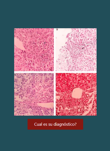
Tumores hepáticos malignos no hepatocelulares. 2.a parte. Aspectos morfológicos y estudios auxiliares de importancia útiles para el diagnóstico histopatológico
DOI:
https://doi.org/10.22516/25007440.63Palabras clave:
Tumores hepáticos malignos, colangiocarcinoma, cistoadenocarcinoma, hemangioendotelioma epitelioide, angiosarcoma, inmunohistoquímicaResumen
Se continúa con la revisión de los tumores malignos hepáticos primarios. Se hablará de los aspectos más importantes de los tumores primarios no hepatocelulares, siendo más frecuentes los originados en el epitelio del ducto biliar como el colangiocarcinoma, el cistoadenocarcinoma y los tumores mesenquimales, y los más infrecuentes el hemangioendotelioma epitelioide y el angiosarcoma. La principal dificultad radica en lograr el diagnóstico definitivo, el cual se basa en la exclusión de una neoplasia primaria extrahepática o de lesiones hepáticas benignas. Estudios adicionales de inmunohistoquímica, estudios de clonalidad o moleculares pue- den ser de mucha utilidad.
Descargas
Lenguajes:
esReferencias bibliográficas
Ustundag Y, Bayraktar Y. Cholangiocarcinoma: A com- pact review of the literature. World J Gastroenterol. 2008;14(42):6458-66.
Mosconi S. Cholangiocarcinoma. Crit Rev Oncol Hemat. 2009;69:259-70.
Rizvi S, Borad MJ, Patel T, Gores GJ. Cholangiocarcinoma: Molecular pathways and therapeutic opportunities. Semin Liver Dis. 2014;34(4):456-64.
Bosman FT, Carneiro F, Hruban RH, Theise ND (editors). WHO Classification or tumours of the digestive system. Lyon, France: IARC; 2010.
Tushar P. Cholangiocarcinoma. Nat Clin Pract Gastroenterol Hepatol. 2006;3(1):33-42.
Shin HR. Epidemiology of cholangiocarcinoma: An update focusing on risk factors. Cancer Sci. 2010;101:579-85.
Villaveces VA, López R. Orthotopic liver transplantation for biliary atresia complicated by incidental cholangiocar- cinoma: Case report and literature review. J Pediatr Gastr Nutr. 2012;55(3):336-7.
Mosconi S. Cholangiocarcinoma. Crit Rev Oncol Hemat. 2009;69:259-70.
Leong T, Wannakrairot P, Lee ES, Leong A. Pathology of cholangiocarcinoma. Curr Diag Path. 2007;13:54-64.
Shaib YH, Davila JA, McGlynn K, et al. Rising incidence of intrahepatic cholangiocarcinoma in the US: A true increase? J Hepatol. 2004;40:472-7.
Shaib Y, El-Serag HB. The epidemiology of cholangiocarci- noma. Sem Liver Dis. 2004;24:115-24.
Goodman ZD. Neoplasms of the liver. Modern Pathol. 2007;20:S49-60.
Liau JY. Morphological subclassification of intrahepatic cholangiocarcinoma: Etiological, clinicopathological, and molecular features. Modern Pathol. 2014;27:1163-73.
Nakanuma Y, Sato Y, Harada K, Sasaki M, Xu J, Ikeda H. Pathological classification of intrahepatic cholangio- carcinoma based on a new concept. World J Hepatol. 2010;2(12):419-27.
Dabbs DJ, Geisinger KR, Ruggiero F, Raab SS, Nalesnik M, Silverman JF. Recommendations for the reporting of tis- sues removed as part of the surgical treatment of malignant liver tumors. The Association of Directors of Anatomic and Surgical Pathology. Am J Clin Pathol. 2005;123:494-8.
Mosnie JF. N-cadherin serves as diagnostic biomarker in intrahepatic and perihilar cholangiocarcinomas. Modern Pathol. 2009;22:182-90.
Zen Y, Pedica F, Patcha VR, Capelli P, Zamboni G, Casaril A, et al. Mucinous cystic neoplasms of the liver: A clinicopatho- logical study and comparison with intraductal papillary neo- plasms of the bile duct. Modern Pathol. 2011;24:1079-89.
Lee CW, Tsai HI, Lin YS, Wu TH, Yu MC, Chen MF. Intrahepatic biliary mucinous cystic neoplasms: Clinicoradiological characteristics and surgical results. BMC Gastroenterol. 2015;15:67-75.
Soares KC, Arnaoutakis DJ, Kamel I, Anders R, Adams RB, Bauer TW, et al. Cystic neoplasms of the liver: biliary cystadenoma and cystadenocarcinoma. J Am Coll Surg. 2014;218(1):119-28.
Baron PW, Amankonah T, Cubas RF, Kore AH, Elihu A, de Vera ME, et al. Diffuse hepatic epithelioid hemangioen- dothelioma developed in a patient with hepatitis C cirrhosis. Case Rep Transplant. 2014;2014:694903.
Zhao XY, Rakhda MI, Habib S, Bihi A, Muhammad A, Wang TL et al. Hepatic epithelioid hemangioendothe- lioma: A comparison of Western and Chinese methods with respect to diagnosis, treatment and outcome. Oncol Lett. 2014;7:977-83.
Campionea S, Cozzolino I, Mainenti P, D’Alessandroa V, Vetrani A, D’Armiento M. Hepatic epithelioid hemangioen- dothelioma: Pitfalls in the diagnosis on fine needle cytology and “small biopsy” and review of the literature. Pathol Res Pract. 2015;211:702-5.
Weinreb I, Cunningham KS, Perez-Ordoñez B, et al. CD10 is expressed in most epitheliod hemangioendotheliomas: A
potential diagnostic pitfall. Arch Pathol Lab Med. 2009;133
(12):1965-8.
Bioulac-Sage P, Laumonier H, Laurent C, et al. Benign and
malignant vascular tumors of the liver in adults. Semin Liver
Dis. 2008;28(3):302-14.
López R, Castro-Villabón D, Álvarez J, Vera A, Andrade R.
Hepatic angiosarcoma presenting as acute liver failure in young adults. Report of two cases and review of literature. Case Reports in Clin Med. 2013;2:439-44.
Srinivasa S, Lee WG, Aldameh A, Koea JB. Spontaneous hepatic hemorrhage: A review of pathogenesis, etiology and treatment. HPB (Oxford). 2015 Aug 7. doi: 10.1111/ hpb.12474.
Deyrup AT, Miettinen M, North PE, Khoury JD, Tighiouart M, et al. Angiosarcomas arising in the viscera and soft tissue of children and young adults. Am J Surg Pathol. 2009;33:264-9.
Wiland HO, Pai RK, Purysko AS. Hepatic angiosarcoma mimicking sinusoidal obstruction syndrome/venoocclusive disease: A pathologic-radiologic correlation. Ann Diagnos Pathol. 2012;16: 275-9.
Zhu YP, Chen YM, Matro E, Chen RB, Jiang ZN, Mou YP, et al. Primary hepatic angiosarcoma: A report of two cases and literature review World J Gastroenterol. 2015;21(19):6088- 96.
Descargas
Publicado
Cómo citar
Número
Sección
Licencia
Aquellos autores/as que tengan publicaciones con esta revista, aceptan los términos siguientes:
Los autores/as ceden sus derechos de autor y garantizarán a la revista el derecho de primera publicación de su obra, el cuál estará simultáneamente sujeto a la Licencia de reconocimiento de Creative Commons que permite a terceros compartir la obra siempre que se indique su autor y su primera publicación en esta revista.
Los contenidos están protegidos bajo una licencia de Creative Commons Reconocimiento-NoComercial-SinObraDerivada 4.0 Internacional.

| Estadísticas de artículo | |
|---|---|
| Vistas de resúmenes | |
| Vistas de PDF | |
| Descargas de PDF | |
| Vistas de HTML | |
| Otras vistas | |