
The usefulness of Giemsa staining to diagnose Helicobacter pylori in patients with preneoplastic lesions
DOI:
https://doi.org/10.22516/25007440.938Keywords:
Histological techniques, Helicobacter pylori, Diagnosis, Preneoplastic lesions, Gastrointestinal diseasesAbstract
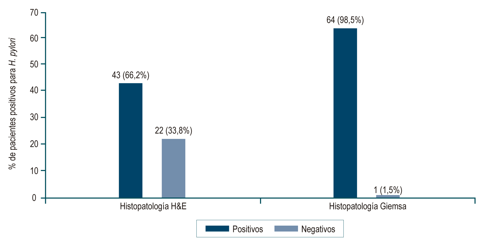
Introduction: Helicobacter pylori is a bacterium associated with inflammatory and neoplastic gastroduodenal diseases. Histopathology is one of the diagnostic methods used for its detection, which has a sensitivity of 90% to 95% when there is a high density of H. pylori; however, the bacterium may be missed in low-density infections because routine hematoxylin and eosin (H&E) staining is not specific for its detection and has interobserver variability. This study aimed to determine the usefulness of complementary Giemsa staining for diagnosing H. pylori in preneoplastic lesions where the bacterium was found in low density.
Materials and methods: A retrospective/prospective descriptive study was carried out that included 65 patients diagnosed with preneoplastic lesions. Gastric biopsies were stained with H&E and Giemsa and evaluated by two pathologists.
Results: Giemsa staining analyzed 20.3% more cases of H. pylori than H&E, most with a low density of the bacteria. There were no statistically significant differences in the diagnosis of H. pylori according to the sample type.
Conclusion: This study found that Giemsa staining improves the histopathological diagnosis of H. pylori in patients with preneoplastic lesions.
Downloads
References
Zamani M, Ebrahimtabar F, Zamani V, Miller WH, Alizadeh-Navaei R, Shokri- Shirvani J, et al. Systematic review with meta-analysis: the worldwide prevalence of Helicobacter pylori infection. Aliment Pharmacol Ther. 2018;47(7):868-76. https://doi.org/10.1111/apt.14561
Roldán IJ, Castaño R, Navas MC. Mutaciones del gen ARN ribosómico 23S de Helicobacter pylori asociadas con resistencia a claritromicina en pacientes atendidos en una unidad de endoscopia de Medellín, Colombia. Biomedica. 2019;39(Supl. 2):117-29. https://doi.org/10.7705/biomedica.v39i4.4377
Bravo LE, Cortés A, Carrascal E, Jaramillo R, García LS, Bravo PE. Helicobacter pylori: patología y prevalencia en biopsias gástricas en Colombia. Colomb Médica. 2003;34(3):124-31.
Correa GS, Cardona A, Correa GT, García G, Estrada S. Prevalencia de Helicobacter pylori y características histopatológicas en biopsias gástricas de pacientes con síntomas dispépticos en un centro de referencia de Medellín. Rev Colomb Gastroenterol. 2016;31(1):9-15. https://doi.org/10.22516/25007440.67
IARC Working Group on the Evaluation of Carcinogenic Risks to Humans. Schistosomes, Liver Flukes and Helicobacter pylori. Lyon (FR): International Agency for Research on Cancer; 1994. (IARC Monographs on the Evaluation of Carcinogenic Risks to Humans, No. 61.) INFECTION WITH HELICOBACTER PYLORI. Available from: https://www.ncbi.nlm.nih.gov/books/NBK487794/
Colombia: source: Globocan 2020 [Internet]. Iarc.fr.; 2021 [citado el 31 de marzo de 2022]. Disponible en: https://gco.iarc.fr/today/data/factsheets/populations/170- colombia-fact-sheets.pdf
Weng C-Y, Xu J-L, Sun S-P, Wang K-J, Lv B. Helicobacter pylori eradication: Exploring its impacts on the gastric mucosa. World J Gastroenterol. 2021;27(31):5152-70. https://doi.org/10.3748/wjg.v27.i31.5152
Basso L, Gallo G, Biacchi D, Carati MV, Cavallaro G, Esposito L, et al. Role of new anatomy, biliopancreatic reflux, and Helicobacter pylori status in postgastrectomy stump cancer. J Clin Med. 2022;11(6):1498. https://doi.org/10.3390/jcm11061498
Bordin DS, Voynovan IN, Andreev DN, Maev IV. Current Helicobacter pylori diagnostics. Diagnostics (Basel). 2021;11(8):1458. https://doi.org/10.3390/diagnostics11081458
Chahuán AJ, La DP, Villalón FA. Métodos de diagnóstico para la detección de la infección por Helicobacter pylori. Rev Gastroenterol Latinoam. 2020;31(2):98-106. https://doi.org/10.46613/gastrolat202002-08
Sabbagh P, Mohammadnia-Afrouzi M, Javanian M, Babazadeh A, Koppolu V, Vasigala VR, et al. Diagnostic methods for Helicobacter pylori infection: ideals, options, and limitations. Eur J Clin Microbiol Infect Dis. 2019;38(1):55-66. https://doi.org/10.1007/s10096-018-3414-4
Nizeyimana T, Rugwizangoga B, Manirakiza F, Laga AC. Occurrence of Helicobacter pylori in specimens of chronic gastritis and gastric adenocarcinoma patients: A retrospective study at university teaching hospital, Kigali, Rwanda. East Afr Health Res J. 2021;5(2):159-63. https://doi.org/10.24248/eahrj.v5i2.667
Zhang C, Yamada N, Wu YL, Wen M, Matsuhisa T, Matsukura N. Helicobacter pylori infection, glandular atrophy and intestinal metaplasia in superficial gastritis, gastric erosion, erosive gastritis, gastric ulcer and early gastric cancer. World J Gastroenterol. 2005;11(6):791-796. https://doi.org/10.3748/wjg.v11.i6.791
Chahuan J, Pizarro M, Riquelme A. Métodos diagnósticos para la detección de infección por Helicobacter pylori. ¿Cuál y cuándo deben solicitarse? Acta Gastroenterol Latinoam. 2022;52(1):36-46. https://doi.org/10.52787/agl.v52i1.176
Loor A, Dumitraşcu DL. Helicobacter pylori Infection, Gastric Cancer and Gastropanel. Rom J Intern Med. 2016;54(3):151-6. https://doi.org/10.1515/rjim-2016-0025
Dixon MF, Genta RM, Yardley JH, Correa P. Classification and grading of gastritis. The updated Sydney System. International Workshop on the Histopathology of Gastritis, Houston 1994. Am J Surg Pathol. 1996;20(10):1161-81. https://doi.org/10.1097/00000478-199610000-00001
Salazar BE, Pérez-Cala T, Gomez-Villegas SI, Cardona-Zapata L, Pazos- Bastidas S, Cardona-Estepa A, et al. The OLGA-OLGIM staging and the interobserver agreement for gastritis and preneoplastic lesion screening: a cross-sectional study. Virchows Arch. 2022;480(4):759-769. https://doi.org/10.1007/s00428-022-03286-8
Mawlood AH, Kawther RS, Balaky S. Evaluation of Invasive and Non-Invasive Methods for the Diagnosis of H. pylori in Dyspepsia Patients. Diyala J Med. 2019;16(2):55-63. https://doi.org/10.26505/DJM.16024460122
Laine L, Lewin DN, Naritoku W, Cohen H. Prospective comparison of H&E, Giemsa, and Genta stains for the diagnosis of Helicobacter pylori. Gastrointest Endosc. 1997;45(6):463-7. https://doi.org/10.1016/S0016-5107(97)70174-3
Miwata T, Quach DT, Hiyama T, Aoki R, Le HM, Tran PLN, et al. Interobserver and intraobserver agreement for gastric mucosa atrophy. BMC Gastroenterol. 2015;15:95. https://doi.org/10.1186/s12876-015-0327-x
Gastritis atrófiaca y Helicobacter pylori. Rev Gastroenterol Peru. 2002;22(3):197-8.
Kong Y-J, Yi H-G, Dai J-C, Wei M-X. Histological changes of gastric mucosa after Helicobacter pylori eradication: a systematic review and meta-analysis. World J Gastroenterol. 2014;20(19):5903-11. https://doi.org/10.3748/wjg.v20.i19.5903
Sung JJY, Coker OO, Chu E, Szeto CH, Luk STY, Lau HCH, et al. Gastric microbes associated with gastric inflammation, atrophy and intestinal metaplasia 1 year after Helicobacter pylori eradication. Gut. 2020;69(9):1572-80. https://doi.org/10.1136/gutjnl-2019-319826
Hwang Y-J, Kim N, Lee HS, Lee JB, Choi YJ, Yoon H, et al. Reversibility of atrophic gastritis and intestinal metaplasia after Helicobacter pylori eradication - a prospective study for up to 10 years. Aliment Pharmacol Ther. 2018;47(3):380-90. https://doi.org/10.1111/apt.14424
Chiang T-H, Chang W-J, Chen SL-S, Yen AM-F, Fann JC-Y, Chiu SY-H, et al. Mass eradication of Helicobacter pylori to reduce gastric cancer incidence and mortality: a long-term cohort study on Matsu Islands. Gut. 2021;70(2):243-50. https://doi.org/10.1136/gutjnl-2020-322200
Shah DK, Jain SS, Mohite A, Amarapurkar AD, Contractor QQ, Rathi PM. Effect of H. pylori density by histopathology on its complications and eradication therapy. Trop Gastroenterol. 2015;36(2):101-6. https://doi.org/10.7869/tg.261
Khan H, Rauf F, Muhammad N, Javaid M, Alam S, Nasir S. Comparación de tinciones especiales (tinción de Giemsa y tinción de azul de toluidina modificada) con inmunohistoquímica como estándar de oro para la detección de H. pylori en biopsias gástricas. Arab J Gastroenterol. 2022;23(2):75-81.
Boldt MS, Pereira RD, Barbosa AJA. Identificación histológica de H. pylori stained por hematoxilina-eosina y Giemsa: revisión para el control de calidad. J Bras Patol Med Lab. 2015;51(2):108-12.
Alkhamiss AS. Evaluation of better staining method among hematoxylin and eosin, Giemsa and periodic acid Schiff-Alcian blue for the detection of Helicobacter pylori in gastric biopsies. Malays J Med Sci. 2020;27(5):53-61. https://doi.org/10.21315/mjms2020.27.5.6
Moayyedi P, Dixon MF. Any role left for invasive tests? Histology in clinical practice. Gut. 1998;43 Suppl 1:S51-5. https://doi.org/10.1136/gut.43.2008.S51
Vaira D, Ricci C, Holton J. Diagnosis of Helicobacter pylori: invasive and non- invasive tests. Best Pract Res Clin Gastroenterol. 2007;21(2):299-313. https://doi.org/10.1016/j.bpg.2006.11.002
Lee JY, Kim N. Diagnosis of Helicobacter pylori by invasive test: histology. Ann Transl Med. 2015;3(1):10. Disponible en: http://dx.doi.org/10.3978/j.issn.2305-5839.2014.11.03
Makristathis A, Hirschl AM, Mégraud F, Bessède E. Review: Diagnosis of Helicobacter pylori infection. Helicobacter. 2019;24 Suppl 1(S1):e12641. https://doi.org/10.1111/hel.12641
Batts KP, Ketover S, Kakar S, Krasinskas AM, Mitchell KA, Wilcox R, et al. Appropriate use of special stains for identifying Helicobacter pylori: Recommendations from the Rodger C. Haggitt Gastrointestinal Pathology Society. Am J Surg Pathol. 2013;37(11):e12-22. https://doi.org/10.1097/PAS.0000000000000097
Sabbagh LC, Otero W, Hani A, Galindo A, Leguízamo A, Maldonado C, et al. Guías de práctica clínica basadas en la evidencia. Guía de práctica clínica para el diagnóstico y tratamiento de la infección por Helicobacter pylori en adultos [Internet]. Asociación Colombiana de Gastroenterología; 2016-2017 [citado el 9 de julio de 2022]. Disponible en: https://www.gastrocol.com/wp-content/uploads/2020/04/GPC3_Helicobacter.pdf
Kocsmár É, Szirtes I, Kramer Z, Szijártó A, Bene L, Buzás GM, et al. Sensitivity of Helicobacter pylori detection by Giemsa staining is poor in comparison with immunohistochemistry and fluorescent in situ hybridization and strongly depends on inflammatory activity. Helicobacter. 2017;22(4):e12387. https://doi.org/10.1111/hel.12387
Downloads
Published
How to Cite
Issue
Section
License

This work is licensed under a Creative Commons Attribution-NonCommercial-NoDerivatives 4.0 International License.
Aquellos autores/as que tengan publicaciones con esta revista, aceptan los términos siguientes:
Los autores/as ceden sus derechos de autor y garantizarán a la revista el derecho de primera publicación de su obra, el cuál estará simultáneamente sujeto a la Licencia de reconocimiento de Creative Commons que permite a terceros compartir la obra siempre que se indique su autor y su primera publicación en esta revista.
Los contenidos están protegidos bajo una licencia de Creative Commons Reconocimiento-NoComercial-SinObraDerivada 4.0 Internacional.

Funding data
-
Universidad de Antioquia
Grant numbers 2014-1062 -
Departamento Administrativo de Ciencia, Tecnología e Innovación (COLCIENCIAS)
Grant numbers 617-2013
| Article metrics | |
|---|---|
| Abstract views | |
| Galley vies | |
| PDF Views | |
| HTML views | |
| Other views | |