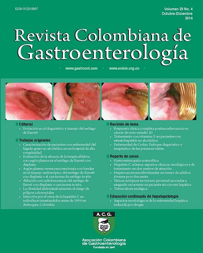
Morphological Issues of Drug Induced Liver Disease
DOI:
https://doi.org/10.22516/25007440.445Keywords:
Liver biopsy, toxicity, drugs, toxins, patterns, necroinflammatory, cholestasis, steatosis, steatohepatitis, green tea, acetaminofén, steroids, aiodarones, methimazole, nitrofurantoin, chlorpromazine, oral contraceptives, methotrexareAbstract
Drug-induced liver disease is a multifaceted phenomenon which has a varied morphological spectrum that mimics other patterns of liver damage both in cases of acute drug exposure and in cases of chronic exposure to drugs. Those patients who are idiosyncratically susceptible at the therapeutic dose or to intrinsic toxicity may also be affected by other factors including genetic factors, age, sex, nutritional status, exposure to other drugs and the existence of an underlying disease. The only clinical manifestation of the disease may be the adverse effect of a drug, but it can also be accompanied by systemic manifestations and manifestations in other organs, and it can even be fatal (1).
The incidence of drug-induced liver disease is not well defined, but some studies claim that its overall annual incidence varies between 1/100,000 people and 15/100,000 people. In the United States, twenty new cases per 100,000 inhabitants occur every year. More than 900 natural and pharmaceutical drugs, herbal medicines, homeopathic products, dietary supplements and toxins have been reported to cause liver damage. This can occur whether or not they are used at normal therapeutic doses. These cases are responsible for about 15% of consultations and hospitalizations for jaundice, acute hepatitis, and chronic hepatitis in adults above the age of 50, and in up to 40% of all cases of hepatitis. Drug-induced liver disease also accounts for 11% to 50% of all cases of acute liver failure. Published data indicate that antibiotics are responsible for between 27% and 46% of cases, that drugs for diseases of the central nervous system are responsible for between 13% and 17%, anti-inflammatory and analgesic agents are responsible for between 5% and 17%, and herbal products are responsible for 9%. New biomarkers and the use of microRNA are being studied and may become promising alternatives in the near future for identifying patients susceptible to drug-induced hepatotoxicity.
There are so many types of liver damage attributed to these agents that only give some examples can be provided in this article. These examples have been chosen on the basis ofn the patterns of liver damage with emphasis on the importance of proper and thorough clinical correlation (2, 3).
Downloads
References
Hou FQ, Zeng Z, Wang GQ. Hospital admissions for drug-induced liver injury: clinical features, therapy, and outcomes. Cell Biochem Biophys 2012; 64: 77-83.
Leise MD, Poterucha JJ, Talwalkar JA. Drug induced liver injury. Mayo Clin Proc 2014; 89(1): 95-106.
Rangnekar AS, Fontana RJ. An update on drug induced liver injury. Minerva Gastroenterol Dietol 2011; 57(2): 213-29.
Ramachandran R, Kakar S. Histological patterns in drug-induced liver disease. J Clin Pathol 2009; 62: 481-92.
Davern TJ. Drug-induced liver disease. Clin Liver Dis 2012; 16: 231-45.
Kleiner DE. The pathology of drug-induced liver injury. Semin Liver Dis 2009; 29: 364-72.
Xuchen Zhang, Jie Ouyang, Swan N. Thung. Histopathologic Manifestations of Drug-induced Hepatotoxicity. Clin Liver Dis 2013; 17: 547-564.
Bjornsson E, Kalaitzakis E, Av Klinteberg V, et al. Long-term follow-up of patients with mild to moderate drug-induced liver injury. Aliment Pharmacol Ther 2007; 26: 79-85.
Andrade RJ, Lucena MI, Kaplowitz N, et al. Outcome of acute idiosyncratic drug-induced liver injury: long-term follow-up in a hepatotoxicity registry. Hepatology 2006; 44: 1581-8.
O’shea, Darasathy, and Mc Cullough Alcoholic Liver Disease. Hepatology 2010; 51(1).
Kleiner DE, Brunt EM. Nonalcoholic fatty liver disease: pathologic patterns and biopsy evaluation in clinical research. Semin Liver Dis 2012; 32: 3-13.
Rocío del Pilar López-P, y col. Ictericia colestásica inducida por metimazol en una paciente con hipertiroidismo. Acta Gastroenterol Latinoam 2014; 44: 52-58.
Abraham C, Hart J, Locke SM and Baker AL. A case of indometacin-induced acute hepatitis developing into chronic autoimmune hepatitis. Gastroenterology & Hepatology 2008; 5(3): 172 -176.
Czaja AJ. Drug-induced autoimmune-like hepatitis. Dig Dis Sci 2011; 56: 958-76.
Suzuki A, Brunt EM, Kleiner DE, et al. The use of liver biopsy evaluation in discrimination of idiopathic autoimmune hepatitis versus drug-induced liver injury. Hepatology 2011; 54: 931-9.
Lewis JH. Diagnosis: liver biopsy differentiates DILI from autoimmune hepatitis. Nat Rev Gastroenterol Hepatol 2011; 8: 540-2.
Andrew S. deLemos, David M. Foureau, Carl Jacobs, Will Ahrens, Mark W. Russo, Herbert L. Bonkovsky. Drug-Induced Liver injury with Autoimmune Features. Semin Liver Dis 2014; 34(02): 194-20.
Gluck N, Fried M and Porat R. Acute Amiodarone Liver Toxicity Likely Due to Ischemic Hepatitis. IMAJ 2011; 748-752.
Mallet VO, Bralet MP, Pol S. Response to Schiano, et al. Hepatoportal sclerosis as a cause of noncirrhotic portal hypertension in patients with HIV. Am J Gastroenterol 2008; 103: 808-9.
Khan AZ, Morris-Stiff G, Makuuchi M. Patterns of chemotherapy-induced hepatic injury and their implications for patients undergoing liver resection for colorectal liver metastases. J Hepatobiliary Pancreat Surg 2009; 16: 137-44.
Teke Z, Nessar G, Kiremitci S, Aksoy E, Elbir O. Hepar Lobatum Carcinomatosum Associated with Metastatic Rectal Carcinoma: An Unusual Cause of Liver Dysmorphy. Med Princ Pract 2011; 20(1): 93-6.
Gravel DH, Bégin LR, Brisson ML, Lamoureux E. Metastatic carcinoma resulting in hepar lobatum. Am J Clin Pathol 1996; 105(5): 621-7.
Infante PF, Petty SE, Groth DH, Markowitz G, Rosner D. Vinyl chloride propellant in hair spray and angiosarcoma of the liver among hairdressers and barbers: case reports. Int J Occup Environ Health 2009; 15(1): 36-42.
Hozo I Miric D, Bojic L, Giunio L, Lusic I, Culic V, Simunic M. Liver angiosarcoma and hemangiopericytoma after occupational exposure to vinyl chloride monomer. Environ Health Perspect 2000; 108(8): 793-5.
Dhingra S, Fiel MI. Update on the new classification of hepatic adenomas: clinical, molecular, and pathologic characteristics. Arch Pathol Lab Med 2014; 138(8): 1090-7.
Daniel F, Cadranel JF, Seksik P, Cazier A, Duong Van Huyen JP, Ziol M, Coutarel P, Loison P, Jian R, Marteau P. Azathioprine induced nodular regenerative hyperplasia in IBD patients. Gastroenterol Clin Biol 2005; 29(5): 600-3.
Bunchorntavakul C, Reddy KR. Herbal and dietary supplement hepatotoxicity. Aliment Pharmacol Ther 2013; 37: 3-17.
Chitturi S, Farrell GC. Corticosteroids. Adverse effects of hormones and hormone antagonists on the liver. En Kaplowitz N, DeLeve LD, eds. Drug-induced liver disease. 3rd ed. Amsterdam: Elsevier; 2013. p. 613-4.
GP Aithal, et al. Monitoring methotrexate-induced hepatic fibrosis in patients with psoriasis: are serial liver biopsies justified? Aliment Pharmacol Ther 2004; 19: 391-399.
MAM Berends, et al. Reliability of the Roenigk Classification of Liver Damage after Methotrexate Treatment for Psoriasis. Arch Dermatol 2007; 143(12): 1515-1519.
Downloads
Published
How to Cite
Issue
Section
License
Aquellos autores/as que tengan publicaciones con esta revista, aceptan los términos siguientes:
Los autores/as ceden sus derechos de autor y garantizarán a la revista el derecho de primera publicación de su obra, el cuál estará simultáneamente sujeto a la Licencia de reconocimiento de Creative Commons que permite a terceros compartir la obra siempre que se indique su autor y su primera publicación en esta revista.
Los contenidos están protegidos bajo una licencia de Creative Commons Reconocimiento-NoComercial-SinObraDerivada 4.0 Internacional.

| Article metrics | |
|---|---|
| Abstract views | |
| Galley vies | |
| PDF Views | |
| HTML views | |
| Other views | |


