Giant esophageal schwannoma, a diagnosis by exclusion: Case report
DOI:
https://doi.org/10.22516/25007440.516Keywords:
Schwannoma, Neurilemoma, Primary esophageal tumor, Esophagus, Case reportAbstract
Introduction: Esophageal schwannomas are tumors of the perineural components of the Schwann cell nerve
sheath in peripheral nerves and account for 2% of primary esophageal tumors. Its low incidence makes
diagnosis challenging; however, this etiology should be considered because its clinical and imaging behavior
is rapidly progressive and unusual compared to other benign esophageal tumors.
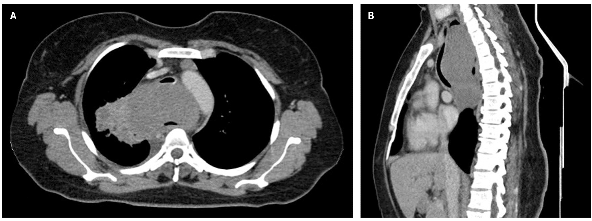
Case study: A 38-year-old female patient with a 1-year history of dysphagia underwent upper digestive tract endoscopy and contrast chest CT showing a mass at the cervical and transmural thoracic esophagus level, obstructing the lumen and exerting a mass effect on the trachea. A biopsy revealed a spindle cell tumor with positive immunohistochemistry for the S100 marker, leading to the diagnosis of esophageal Schwannoma. The patient is currently undergoing regular check-ups and is awaiting the advice of a clinical oncologist to recommend non-surgical treatment options due to the involvement of adjacent structures.
Conclusion: The first diagnostic impression in the case of a suspected primary esophageal tumor would be a leiomyoma based on its incidence. The present case report of an esophageal schwannoma emphasizes that this is a differential diagnosis that requires timely treatment to avoid complications and sequelae in patients.
Downloads
References
Brunicardi FC, Billiar TR, Dunn DL, Hunter JG, Matthews JB, Pollock RE, Wu JX. Schwartz. Principios de cirugía. Autoevaluación y repaso. 10.a edición. Mc-Graw Hill; 2010.
Kitada M, Matsuda Y, Hayashi S, Ishibashi K, Oikawa K, Miyokawa N. Esophageal schwannoma: a case report. World J Surg Oncol. 2013;11:253. https://doi.org/10.1186/1477-7819-11-253
Zhu L, Li W, Zhu Z, Chai Y. Benign esophageal schwannoma: A case report and review of literature. Niger J Clin Pract. 2019;22(5):731-733. https://doi.org/10.4103/njcp.njcp_142_18
Kobayashi N, Kikuchi S, Shimao H, Hiki Y, Kakita A, Mitomi H, Ohbu M. Benign esophageal schwannoma: report of a case. Surg Today. 2000;30(6):526-9. https://doi.org/10.1007/s005950070120
Morales-Maza J, Pastor-Sifuentes FU, Sánchez-Morales GE, Ramos ES, Santes O, Clemente-Gutiérrez U, Pimienta-Ibarra AS, Medina-Franco H. Clinical characteristics and surgical treatment of schwannomas of the esophagus and stomach: A case series and systematic review. World J Gastrointest Oncol. 2019;11(9):750-760. https://doi.org/10.4251/wjgo.v11.i9.750
Souza LCA, Pinto TDA, Cavalcanti HOF, Rezende AR, Nicoletti ALA, Leão CM, Cunha VC. Esophageal schwannoma: Case report and epidemiological, clinical, surgical and immunopathological analysis. Int J Surg Case Rep. 2019;55:69-75. https://doi.org/10.1016/j.ijscr.2018.10.084
Souza LCA, Pinto TDA, Cavalcanti HOF, Rezende AR, Nicoletti ALA, Leão CM, Cunha VC. Esophageal schwannoma: Case report and epidemiological, clinical, surgical and immunopathological analysis. Int J Surg Case Rep. 2019;55:69-75. https://doi.org/10.1016/j.ijscr.2018.10.084
Zhang Y, Han Y, Xiang J, Li H. Robot-assisted enucleation of large dumbbell-shaped esophageal schwannoma: a case report. BMC Surg. 2018;18(1):36. https://doi.org/10.1186/s12893-018-0370-y
Quintero Rivera JC, Arias Morales Y, Carcacía Hermilla I, Prieto Casal PL, Armesto Fernandez MJ, Ourense ES. Schwannomas extracraneales. Hallazgos radiológicos y diagnóstico diferencial. Congreso Nacional de la Sociedad de Radiología Española 2012. Granada; 2012.
Park BJ, Carrasquillo J, Bains MS, Flores RM. Giant benign esophageal schwannoma requiring esophagectomy. Ann Thorac Surg. 2006;82(1):340-2. https://doi.org/10.1016/j.athoracsur.2005.09.042
Wang S, Zheng J, Ruan Z, Huang H, Yang Z, Zheng J. Long-term survival in a rare case of malignant esophageal schwannoma cured by surgical excision. Ann Thorac Surg. 2011;92(1):357-8. https://doi.org/10.1016/j.athoracsur.2011.01.045
Mishra B, Madhusudhan KS, Kilambi R, Das P, Pal S, Srivastava DN. Malignant Schwannoma of the Esophagus: A Rare Case Report. Korean J Thorac Cardiovasc Surg. 2016;49(1):63-6. https://doi.org/10.5090/kjtcs.2016.49.1.63
Walker C, Chung J. Muller’s Imaging of the Chest. 2.a edición. Elsevier; 2018.
Downloads
Published
How to Cite
Issue
Section
License
Aquellos autores/as que tengan publicaciones con esta revista, aceptan los términos siguientes:
Los autores/as ceden sus derechos de autor y garantizarán a la revista el derecho de primera publicación de su obra, el cuál estará simultáneamente sujeto a la Licencia de reconocimiento de Creative Commons que permite a terceros compartir la obra siempre que se indique su autor y su primera publicación en esta revista.
Los contenidos están protegidos bajo una licencia de Creative Commons Reconocimiento-NoComercial-SinObraDerivada 4.0 Internacional.

| Article metrics | |
|---|---|
| Abstract views | |
| Galley vies | |
| PDF Views | |
| HTML views | |
| Other views | |