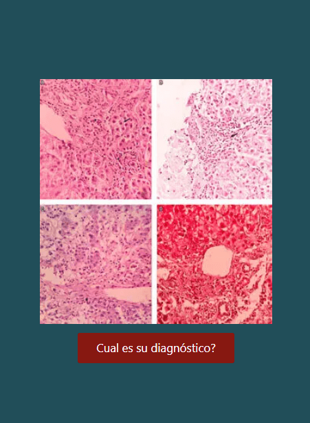
Aspectos morfológicos de la enfermedad hepática inducida por drogas
DOI:
https://doi.org/10.22516/25007440.445Palabras clave:
hígado, biopsia, fármacos, toxicidad, drogas, tóxicos, patrones, necroinflamatorio, colestasis, esteatosis, esteatohepatitis, té verde, acetaminofén, esteroides, amiodarona, metimazol, clorpromacina, anticonceptivos orales, metrotrexateResumen
La enfermedad hepática inducida por drogas es un fenómeno multifacético y su espectro morfológico es muy variado imitando cualquier patrón de daño hepático, tanto en pacientes expuestos en forma aguda o crónica, en aquellos susceptibles en forma idiosincrática a una dosis terapéutica o por toxicidad intrínseca, a su vez puede estar afectada por otros factores como son los genéticos, la edad o el sexo, el estado nutricional, la exposición a otros fármacos o la existencia de una enfermedad de base; puede ser la única manifestación clínica del efecto adverso de una droga o estar acompañado de manifestaciones sistémicas o de otros órganos, e incluso puede llegar a ser fatal (1).
Su incidencia no está bien definida, algunos estudios afirman que la incidencia global es variable encontrándose entre 1-15 x 100.000 personas/año, en USA ocurren 20 nuevos casos x 100.000 habitantes/año. Se han descrito como causantes de lesión hepática más de 900 drogas, productos herbales, homeopáticos, suplementos dietéticos o toxinas, sean productos naturales o de la industria farmacéutica, utilizados o no en dosis terapéuticas, que son las responsables de aproximadamente 15% de consultas y hospitalizaciones por ictericia, hepatitis aguda o crónica; en la población adulta, por encima de los 50 años, llega a 40% de todos los casos de hepatitis. Es también la causante de 11-50% de casos de falla hepática aguda. Los datos publicados indican que los antibióticos son responsables entre un 27-46% de los casos, seguidos por medicamentos para enfermedades del sistema nervioso central entre 13-17%, antiinflamatorios y analgésicos de 5-17% y los productos herbales 9%. Nuevos biomarcadores y el uso de microRNA se están estudiando y serán prometedores en un futuro cercano para identificar pacientes que puedan presentar hepatotoxicidad inducida por medicamentos.
Son tantos los tipos de lesión hepática atribuidos a estos agentes que solo podremos dar algunos ejemplos en este artículo, basados en los patrones de daño hepático y enfatizando la importancia de una adecuada y profunda correlación clínica (2, 3).
Descargas
Referencias bibliográficas
Hou FQ, Zeng Z, Wang GQ. Hospital admissions for drug-induced liver injury: clinical features, therapy, and outcomes. Cell Biochem Biophys 2012; 64: 77-83.
Leise MD, Poterucha JJ, Talwalkar JA. Drug induced liver injury. Mayo Clin Proc 2014; 89(1): 95-106.
Rangnekar AS, Fontana RJ. An update on drug induced liver injury. Minerva Gastroenterol Dietol 2011; 57(2): 213-29.
Ramachandran R, Kakar S. Histological patterns in drug-induced liver disease. J Clin Pathol 2009; 62: 481-92.
Davern TJ. Drug-induced liver disease. Clin Liver Dis 2012; 16: 231-45.
Kleiner DE. The pathology of drug-induced liver injury. Semin Liver Dis 2009; 29: 364-72.
Xuchen Zhang, Jie Ouyang, Swan N. Thung. Histopathologic Manifestations of Drug-induced Hepatotoxicity. Clin Liver Dis 2013; 17: 547-564.
Bjornsson E, Kalaitzakis E, Av Klinteberg V, et al. Long-term follow-up of patients with mild to moderate drug-induced liver injury. Aliment Pharmacol Ther 2007; 26: 79-85.
Andrade RJ, Lucena MI, Kaplowitz N, et al. Outcome of acute idiosyncratic drug-induced liver injury: long-term follow-up in a hepatotoxicity registry. Hepatology 2006; 44: 1581-8.
O’shea, Darasathy, and Mc Cullough Alcoholic Liver Disease. Hepatology 2010; 51(1).
Kleiner DE, Brunt EM. Nonalcoholic fatty liver disease: pathologic patterns and biopsy evaluation in clinical research. Semin Liver Dis 2012; 32: 3-13.
Rocío del Pilar López-P, y col. Ictericia colestásica inducida por metimazol en una paciente con hipertiroidismo. Acta Gastroenterol Latinoam 2014; 44: 52-58.
Abraham C, Hart J, Locke SM and Baker AL. A case of indometacin-induced acute hepatitis developing into chronic autoimmune hepatitis. Gastroenterology & Hepatology 2008; 5(3): 172 -176.
Czaja AJ. Drug-induced autoimmune-like hepatitis. Dig Dis Sci 2011; 56: 958-76.
Suzuki A, Brunt EM, Kleiner DE, et al. The use of liver biopsy evaluation in discrimination of idiopathic autoimmune hepatitis versus drug-induced liver injury. Hepatology 2011; 54: 931-9.
Lewis JH. Diagnosis: liver biopsy differentiates DILI from autoimmune hepatitis. Nat Rev Gastroenterol Hepatol 2011; 8: 540-2.
Andrew S. deLemos, David M. Foureau, Carl Jacobs, Will Ahrens, Mark W. Russo, Herbert L. Bonkovsky. Drug-Induced Liver injury with Autoimmune Features. Semin Liver Dis 2014; 34(02): 194-20.
Gluck N, Fried M and Porat R. Acute Amiodarone Liver Toxicity Likely Due to Ischemic Hepatitis. IMAJ 2011; 748-752.
Mallet VO, Bralet MP, Pol S. Response to Schiano, et al. Hepatoportal sclerosis as a cause of noncirrhotic portal hypertension in patients with HIV. Am J Gastroenterol 2008; 103: 808-9.
Khan AZ, Morris-Stiff G, Makuuchi M. Patterns of chemotherapy-induced hepatic injury and their implications for patients undergoing liver resection for colorectal liver metastases. J Hepatobiliary Pancreat Surg 2009; 16: 137-44.
Teke Z, Nessar G, Kiremitci S, Aksoy E, Elbir O. Hepar Lobatum Carcinomatosum Associated with Metastatic Rectal Carcinoma: An Unusual Cause of Liver Dysmorphy. Med Princ Pract 2011; 20(1): 93-6.
Gravel DH, Bégin LR, Brisson ML, Lamoureux E. Metastatic carcinoma resulting in hepar lobatum. Am J Clin Pathol 1996; 105(5): 621-7.
Infante PF, Petty SE, Groth DH, Markowitz G, Rosner D. Vinyl chloride propellant in hair spray and angiosarcoma of the liver among hairdressers and barbers: case reports. Int J Occup Environ Health 2009; 15(1): 36-42.
Hozo I Miric D, Bojic L, Giunio L, Lusic I, Culic V, Simunic M. Liver angiosarcoma and hemangiopericytoma after occupational exposure to vinyl chloride monomer. Environ Health Perspect 2000; 108(8): 793-5.
Dhingra S, Fiel MI. Update on the new classification of hepatic adenomas: clinical, molecular, and pathologic characteristics. Arch Pathol Lab Med 2014; 138(8): 1090-7.
Daniel F, Cadranel JF, Seksik P, Cazier A, Duong Van Huyen JP, Ziol M, Coutarel P, Loison P, Jian R, Marteau P. Azathioprine induced nodular regenerative hyperplasia in IBD patients. Gastroenterol Clin Biol 2005; 29(5): 600-3.
Bunchorntavakul C, Reddy KR. Herbal and dietary supplement hepatotoxicity. Aliment Pharmacol Ther 2013; 37: 3-17.
Chitturi S, Farrell GC. Corticosteroids. Adverse effects of hormones and hormone antagonists on the liver. En Kaplowitz N, DeLeve LD, eds. Drug-induced liver disease. 3rd ed. Amsterdam: Elsevier; 2013. p. 613-4.
GP Aithal, et al. Monitoring methotrexate-induced hepatic fibrosis in patients with psoriasis: are serial liver biopsies justified? Aliment Pharmacol Ther 2004; 19: 391-399.
MAM Berends, et al. Reliability of the Roenigk Classification of Liver Damage after Methotrexate Treatment for Psoriasis. Arch Dermatol 2007; 143(12): 1515-1519.
Descargas
Publicado
Cómo citar
Número
Sección
Licencia
Aquellos autores/as que tengan publicaciones con esta revista, aceptan los términos siguientes:
Los autores/as ceden sus derechos de autor y garantizarán a la revista el derecho de primera publicación de su obra, el cuál estará simultáneamente sujeto a la Licencia de reconocimiento de Creative Commons que permite a terceros compartir la obra siempre que se indique su autor y su primera publicación en esta revista.
Los contenidos están protegidos bajo una licencia de Creative Commons Reconocimiento-NoComercial-SinObraDerivada 4.0 Internacional.

| Estadísticas de artículo | |
|---|---|
| Vistas de resúmenes | |
| Vistas de PDF | |
| Descargas de PDF | |
| Vistas de HTML | |
| Otras vistas | |